
The Immun-Star WesternC kit is a complementary chemiluminescent detection kit that is optimised for CCD imaging of the WesternC standards and protein samples.Ĭurrently, Bio-Rad offers a sample size of both the WesternC standard and the Immun-Star WesternC kit.īio-Rad envisions that the Precision Plus Protein WesternC standard will become the leading western blotting standard in all areas of biological research. The Precision Plus Protein WesternC standards offers ten prestained protein bands that can also be detected through chemiluminescent western blotting. The membrane is immediately scanned using an imager following manufacturer’s instructions.Bio-Rad Laboratories has announced the release of a broad-range, prestained chemiluminescent protein standard and a chemiluminescent detection kit for protein sizing and expression analysis.The antibodies are visualized by mixing the chemiluminescence development substrates, Peroxide Buffer and Luminol/Enhancer Solution (Pierce), in a 1:1 ratio and incubating this mixture on the membrane for 5 minutes.For chemiluminescence detection, the secondary antibody, horseradish peroxidase-conjugated α-mouse IgG, is incubated with the membrane at a 1:10,000 dilution in TBST for 1 hour.The reactions are terminated by washing the membranes in water. The antibodies are visualized by adding the color development substrates, Nitro blue tetrazolium (NBT) and 5-bromo-4-chloro-3-indolylphosphate (BCIP), in alkaline phosphatase buffer (100 mM NaCl, 5 mM MgCl2, 100 mM Tris-HCl, pH 9.5).An additional 30 second wash with TBS is carried out.
#Bio rad western blot all blue standards free#
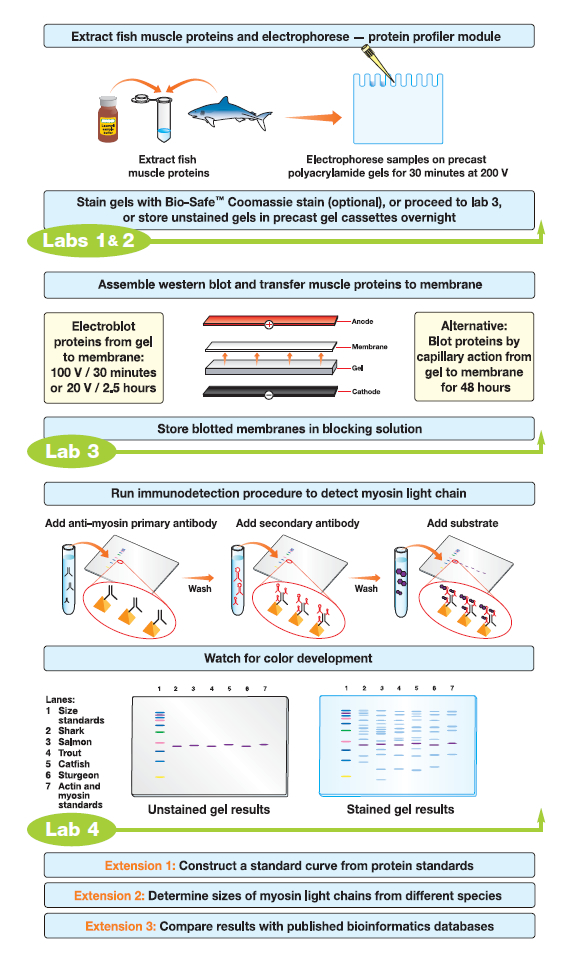
The membrane is washed with TBST as before to remove any free secondary antibody.For colorimetric detection, the secondary antibody alkaline, phosphataseconjugated α-rabbit IgG, is incubated with the membrane at appropriate dilution in TBST for 1 hour at room temperature.The membranes are washed 3 times with TBST for 10 minutes remove free primary antibody.The primary antibody is used following the manufacturer’s advice. The membranes are incubated in appropriate dilutions of primary antibody in TBST (150 mM NaCl, 0.05% Tween 20, 20 mM Tris-HCl, pH 7.5) for 1 hour at room temperature or overnight at 4☌, with carefully shaking.After transfer, the membranes are blocked with a BSA blocking solution (5% BSA, 150 mM NaCl, 0.05% Tween 20, 20 mM Tris-HCl, pH 7.5) or a milk blocking solution (5% powdered milk, 150 mM NaCl, 0.05% Tween 20, 20 mM Tris-HCl, pH 7.5).All of the remaining steps are carried out at room temperature. The protein transfer is carried out at 100 V for 1 hour at 4☌ with buffer recirculation. The transfer is carried out in transfer buffer (192 mM glycine, 20% v/v methanol, 25 mM Tris-HCl, pH 8.3). Proteins fractionated by SDS-PAGE are electrotransferred onto polyvinylidene difluoride (PVDF) membranes.The Mini Trans-Blot Electrophoretic Transfer Cell system (Bio-Rad) is used for western blot experiments.Gels are either electrotransferred to PVDF membranes.Gels are electrophoresed between 100 and 150 V in running buffer (3% Tris-HCl, 14.4% glycine, 1% SDS) until the bromophenol blue dye reaches the bottom of the gel.Samples are loaded and fractionated on 8, 10, or 12% polyacrylamide gels along with a protein ladder.SDS-PAGE is performed using the Mini-PROTEAN III electrophoresis system (Bio-Rad). The standard curve is produced by using varying concentrations of bovine serum albumin (BSA). The reading obtained is compared to a standard curve that allowed for a relative measurement of protein concentration. The Bradford assay method (Bio-Rad Protein Assay) is utilized to determine the concentration of proteins in each sample. Usually, samples are resuspended or mixed in 20-50 µl of 1X sample buffer and all the mixtures are incubated at 90☌ for five minutes or 50☌ for 15 minutes to break down the interactions between peptides. The schematic diagram of western bolt protocol.


 0 kommentar(er)
